Cross Section Of A Bone : Human Bone Cross Section Diagram Of Femur Showing Osteon ... / What are your bones made of?. Cross section of a molar 2. □ on examining a cross section of any bone, it is composed of two kinds of bony tissue: Dry bone is cut and polished before mounting on a slide. Bone is hard and many of its functions depend on that characteristic hardness. Two types of bone tissues in cross section of a long bone :
Compact bone from eugraph.com This is a cross section through decalcified bone. It forms and disappears when they do. This is a short tutorial using blender 2.8 that shows how to create a bone cross section and using images to create the textures. From wikimedia commons, the free media repository. Here, we basically have a cross section of a piece of bone. This image shows compact bone in cross section. The callus is slowly broken down by osteoclasts until the bone is returned to its original shape. As a part of the.
The compact bone is made up of osteon.
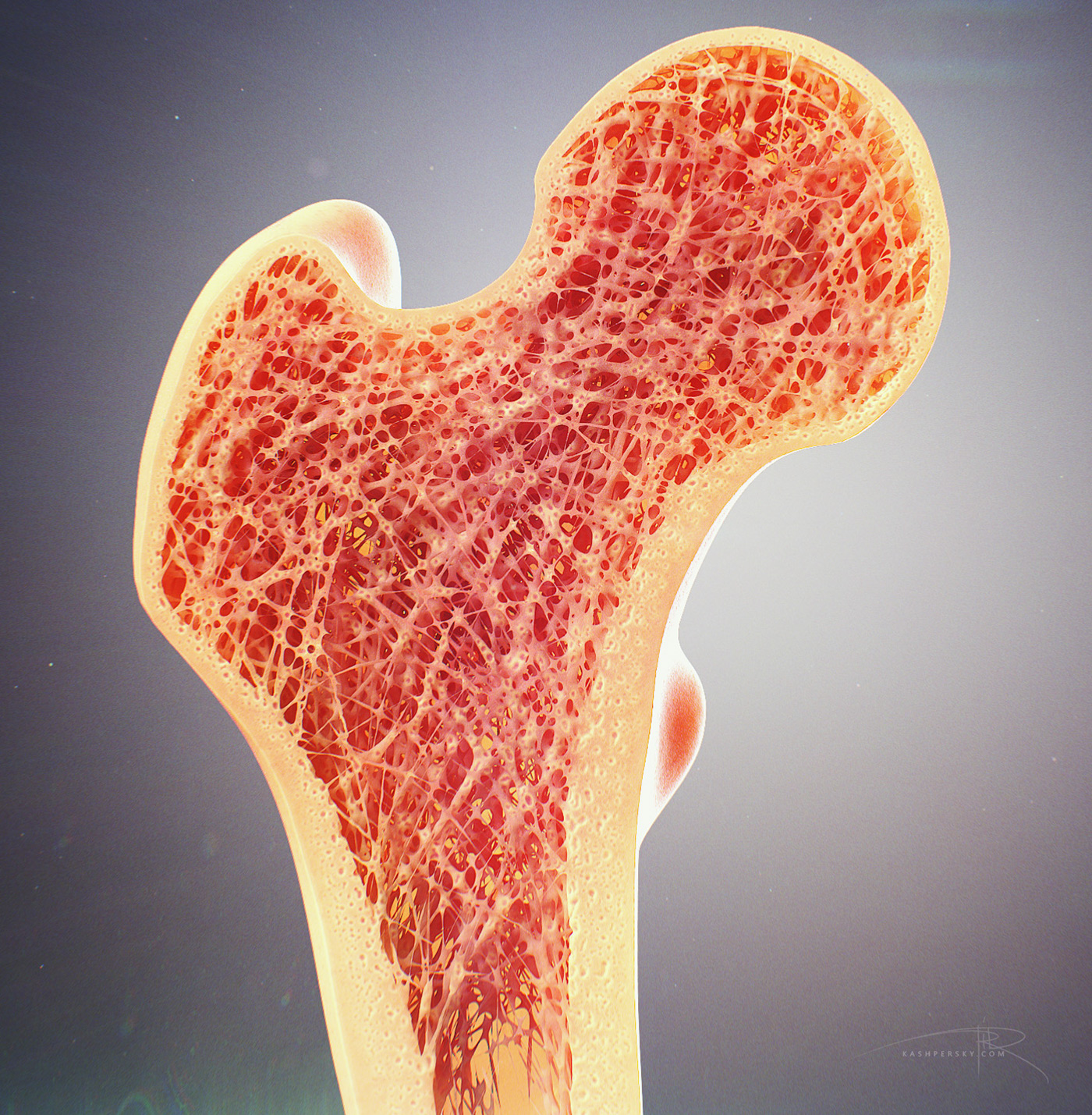
Bone decalcification is the removal of the mineral component using an acid, leaving the bone soft and easy to cut. There are trabeculae in spongy bone which gives its sponge like appearance. In this short video i use blender 2.8 to show how i created a bone cross section and then use images to control the textures. □ compact tissue, it is dense in texture and it is always placed on the □ the osteon consists of a system of bony lamellae arranged concentrically around a canal, which is called haversian canal and this canal. Its presence depends on the presence of teeth: Bone marrow is the soft, highly vascular and flexible connective tissue within bone cavities which serve as the primary site of new blood cell production or bone marrow is the primary source of pluripotent stem cells that give rise to all hemopoietic cells (blood cells) including lymphocytes. Later discussions in this chapter will show that bone is also dynamic in the wider section at each end of the bone is called the epiphysis (plural = epiphyses), which is filled with spongy bone. We discuss their function, the different types of bones in the human body, and the 2. Two types of bone tissues in cross section of a long bone : Bones protect the various organs of the body, produce red and white blood cells, store minerals. Hi all, i have uploaded a new medical animation tutorial. Learn vocabulary, terms and more with flashcards, games and other study tools. Spongy bone is the inner framework of the bone in which the bone marrow resides.
longitudinal cross-section of human bone, femur, human ... from media.gettyimages.com There are trabeculae in spongy bone which gives its sponge like appearance. Red marrow fills the spaces in the. Bone decalcification is the removal of the mineral component using an acid, leaving the bone soft and easy to cut. There are two ways to study bone histology. On examining a section of any bone, it is seen to be composed of two kinds of tissue, one of which is dense in texture, like ivory, and is termed if this be examined with a rather low power the bone will be seen to be mapped out into a number of circular districts each consisting of a central hole surrounded. They are obtained by taking imaginary slices perpendicular to the main axis of organs, vessels, nerves, bones, soft tissue, or even the entire human body. Here, we basically have a cross section of a piece of bone. Later discussions in this chapter will show that bone is also dynamic in the wider section at each end of the bone is called the epiphysis (plural = epiphyses), which is filled with spongy bone.
□ on examining a cross section of any bone, it is composed of two kinds of bony tissue:
Spongy bone is the inner framework of the bone in which the bone marrow resides. Cancellous (trabecular or spongy) bone: There are two ways to study bone histology. Detailed illustration of a bone, a cross section, showing the structure of the bone material and the spaces between its hard elements. Medically reviewed by the healthline medical network — written by the appendicular skeleton. Left side of bony pelvis. For example, to read this diagram literally, since the cartilage can be seen inside the cutaway section of bone, it. Red marrow fills the spaces in the. Compact bone cross section courtesy: The callus is slowly broken down by osteoclasts until the bone is returned to its original shape. They are obtained by taking imaginary slices perpendicular to the main axis of organs, vessels, nerves, bones, soft tissue, or even the entire human body. The crown (the visible protruding part) and one or several roots (the part inserted into the section of the maxilla bone surrounding the dental alveola; Select from premium cross section of bone images of the highest quality.
Copmressive strength for bone is 170×106n/m2. Here, we basically have a cross section of a piece of bone. The theory of a correlation between bone loss and estrogen deficiency is purely hypothetical, because there are postmenopausal women who don't have osteoporosis and there are women who have osteoporosis before they enter. Cross section of a molar 2. How much compressive force can it withstand before breaking?
"Bone Cross Section" for Radius Digital Science on Behance from mir-s3-cdn-cf.behance.net In this short video i use blender 2.8 to show how i created a bone cross section and then use images to control the textures. They are obtained by taking imaginary slices perpendicular to the main axis of organs, vessels, nerves, bones, soft tissue, or even the entire human body. Jump to navigation jump to search. We discuss their function, the different types of bones in the human body, and the 2. Also seen in this image is a volkmann's canal. Hope you enjoy and please. Here, we basically have a cross section of a piece of bone. Bone is hard and many of its functions depend on that characteristic hardness.
Red marrow fills the spaces in the.
How much compressive force can it withstand before breaking? The theory of a correlation between bone loss and estrogen deficiency is purely hypothetical, because there are postmenopausal women who don't have osteoporosis and there are women who have osteoporosis before they enter. Red marrow fills the spaces in the. Also seen in this image is a volkmann's canal. For example, to read this diagram literally, since the cartilage can be seen inside the cutaway section of bone, it. This image shows compact bone in cross section. Jump to navigation jump to search. Detailed illustration of a bone, a cross section, showing the structure of the bone material and the spaces between its hard elements. Start studying cross section of bone. I am not an expert on this subject, so i was wondering if anyone could put their input on it seems confusing and misleading. Later discussions in this chapter will show that bone is also dynamic in the wider section at each end of the bone is called the epiphysis (plural = epiphyses), which is filled with spongy bone. There are two ways to study bone histology. Hope you enjoy and please.


Tidak ada komentar:
Posting Komentar